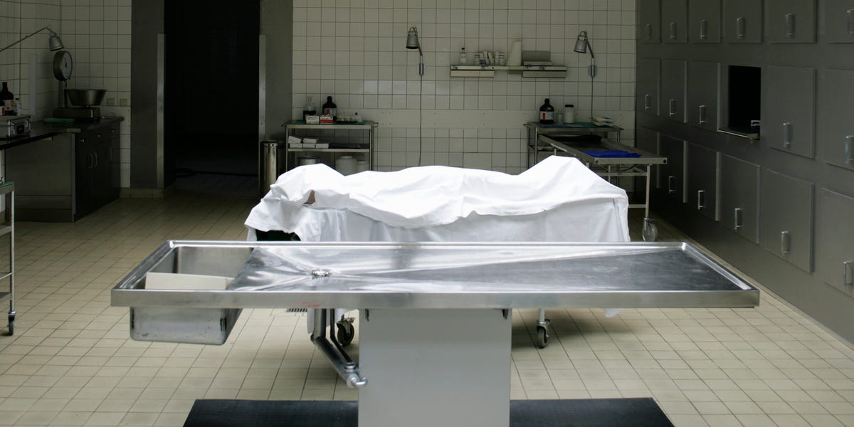

Long before CT scanners, blood tests, and doctors who could casually say, “Let’s get an MRI,” medicine had a stubborn problem: the human body kept its secrets remarkably well. Symptoms could be observed, treatments could be attempted, and theories could be argued over for centuriesbut the decisive evidence was often hidden beneath the skin.
The history of the autopsy is the story of physicians learning to look for themselves. It stretches from ancient anatomical investigations and medieval university dissections to Renaissance pathology, nineteenth-century microscopes, modern medical examiner offices, and today’s virtual autopsy technology. Along the way, postmortem examinations corrected medical myths, exposed crimes, improved public health, and occasionally revealed that a very confident doctor had been confidently wrong.
What Is an Autopsy?
An autopsy is a systematic medical examination of a body after death. A pathologist may inspect the outside of the body, examine internal organs, collect tissue for microscopic analysis, and order toxicology, microbiology, genetic, or imaging tests. The purpose may be to identify disease, determine the cause of death, evaluate medical treatment, investigate an unexpected death, or contribute to scientific research.
The word autopsy comes from the Greek autopsia, meaning “seeing with one’s own eyes” or “seeing for oneself.” That definition captures the procedure’s historical importance. An autopsy replaces assumption with observationor, at the very least, gives assumption a stern performance review.
Clinical Autopsy vs. Forensic Autopsy
A clinical or hospital autopsy generally focuses on disease and medical care. It may confirm a diagnosis, identify an inherited disorder, reveal an unexpected complication, or show why treatment did not work. A forensic autopsy addresses legal and public-interest questions, especially when death is sudden, violent, suspicious, unexplained, or related to an accident.
Forensic pathologists do more than examine organs. They integrate autopsy findings with medical records, death-scene evidence, witness accounts, toxicology results, and other investigative information. The body is one chapter of the casenot the entire book.
Ancient Beginnings: Curiosity Meets Cultural Limits
Ancient civilizations possessed considerable knowledge of injuries, embalming, and internal anatomy, but that knowledge did not automatically produce systematic medical autopsies. Egyptian embalmers handled internal organs, for example, yet their work was primarily religious and funerary rather than an organized investigation of disease.
Greek physicians initially learned much of their anatomy from wounds, animal dissection, and external observation. Hippocratic medicine shifted attention toward natural explanations for illness, but the humoral theory of disease still dominated: health supposedly depended on the balance of blood, phlegm, yellow bile, and black bile. It was an elegant system, provided nobody asked the liver to confirm it.
Herophilus and Erasistratus
A major turning point occurred in Alexandria during the third century BCE. Herophilus and Erasistratus are associated with some of the earliest documented systematic dissections of human bodies. Herophilus studied the brain, nerves, eyes, blood vessels, and reproductive organs, while Erasistratus investigated the heart and vascular system.
Their work demonstrated how direct examination could correct inherited beliefs. However, routine human dissection did not continue. Religious concerns, social discomfort, legal restrictions, and respect for the dead limited access to human bodies across much of the ancient world.
Galen and the Long Rule of Animal Anatomy
In the second century CE, the physician Galen created an enormously influential system of anatomy and medicine. Because human dissection was generally unavailable to him, he relied heavily on animals, including monkeys and pigs. Some structures resembled human anatomy; others did not.
Galen’s writings became so authoritative that later scholars often treated them as settled truth. For more than a millennium, a conflict between Galen’s text and a human body might be blamed on the body. Apparently, even anatomy could be accused of not reading the textbook.
Medieval Autopsies Were Rarer, Not Completely Forbidden
Popular retellings often claim that medieval Europe universally prohibited dissection. The real history is more complicated. Human dissection was limited and controversial, but it was not absent. Postmortem examinations sometimes took place for legal investigations, embalming, religious inquiries, and medical teaching.
Authorities occasionally requested examinations to determine whether poisoning, injury, or disease caused a death. These cases were not modern autopsies with standardized reports and laboratory testing, but they established an important principle: internal findings could answer questions that witnesses and speculation could not.
Mondino de Luzzi and the Return of Public Dissection
In 1315, the Italian physician Mondino de Luzzi conducted a well-documented public human dissection at Bologna, probably involving the body of an executed woman. His anatomical manual, completed around 1316, became an influential guide for university instruction.
Mondino’s dissections were largely intended to teach anatomy rather than investigate disease. Even so, they helped restore direct examination of the human body to European medicine. In the university anatomy theater, the body gradually became evidence rather than merely an illustration of ancient authority.
The Renaissance: Anatomy Challenges the Old Books
Renaissance scholars increasingly valued observation, measurement, and firsthand investigation. Artists wanted accurate muscles. Surgeons wanted accurate organs. Physicians wanted explanations that worked outside a philosophical debate. Human dissection became more regular in major European medical centers, although access to bodies remained ethically and socially fraught.
Antonio Benivieni Connects Symptoms With Findings
Florentine physician Antonio Benivieni recorded detailed clinical cases during the late fifteenth century and sometimes compared symptoms observed during life with findings after death. His collection was published posthumously in 1507. Of the 111 published cases, approximately 16 included postmortem reports.
That number may sound modest, but the method was revolutionary. Benivieni was not opening bodies merely to demonstrate where the spleen lived. He was asking why a particular patient had suffered and died. His observations included intestinal obstruction, gallstones, abscesses, tumors, and other abnormalities that offered anatomical explanations for clinical symptoms.
Vesalius Corrects Galen
Andreas Vesalius transformed anatomical science with the publication of De Humani Corporis Fabrica in 1543. By personally studying human bodies, he identified errors inherited from Galen’s animal-based anatomy. Vesalius did not create modern pathological autopsy by himself, but he established a habit essential to its development: trust careful observation more than reputation.
His work also improved anatomical illustration, teaching, and dissection technique. Medicine was slowly moving from “Galen says” to “the evidence shows,” a change that undoubtedly made faculty meetings much more interesting.
From Collections of Cases to Modern Pathology
Théophile Bonet Builds a Library of the Dead
In 1679, Swiss physician Théophile Bonet published Sepulchretum, a massive compilation of postmortem observations drawn from centuries of medical literature. The work organized thousands of case descriptions by disease and body region, creating one of the earliest systematic reference collections in pathological anatomy.
Bonet’s achievement was largely organizational rather than experimental. He assembled scattered observations so physicians could compare cases. In modern terms, he created a databaseonly with heavier books, more Latin, and absolutely no “forgot password” button.
Giovanni Battista Morgagni Finds the Seat of Disease
The decisive breakthrough came with Giovanni Battista Morgagni. In 1761, at age 79, he published De Sedibus et Causis Morborum per Anatomen Indagatis, commonly translated as On the Seats and Causes of Diseases Investigated by Anatomy.
Morgagni connected detailed medical histories with findings from hundreds of postmortem examinations. Instead of attributing illness mainly to an invisible imbalance of humors, he argued that disease could be located in particular organs and anatomical lesions. Chest pain, paralysis, coughing, vomiting, and other symptoms could be traced to structural changes within the body.
This clinicopathological method became a foundation of modern medicine: observe the patient during life, examine the body after death, and compare the two. The autopsy was no longer a dramatic final inspection. It became a method for testing diagnosis itself.
The Nineteenth-Century Golden Age of Autopsy
During the nineteenth century, hospitals expanded, medical records improved, and pathology emerged as a separate discipline. Autopsies became central to medical education and hospital research. Physicians could compare many patients with similar symptoms, identify recurring lesions, and classify diseases more accurately.
Xavier Bichat Moves From Organs to Tissues
French anatomist Xavier Bichat argued that organs were composed of distinct tissues and that disease could affect those tissues differently. Although he worked before modern microscopy became routine, his tissue-based approach refined Morgagni’s organ-centered model.
The medical focus had moved from the whole person’s humors to organs, then from organs to tissues. The next stop would be the cell.
Karl von Rokitansky Standardizes the Examination
At Vienna General Hospital, Karl von Rokitansky helped turn autopsy into a systematic institutional practice. He is traditionally credited with personally performing approximately 30,000 autopsies and supervising tens of thousands more. The extraordinary volume allowed him to recognize patterns that might remain invisible in isolated cases.
Rokitansky improved gross pathological description and encouraged consistent examination of the organs. His work made the autopsy a central engine of hospital medicine, although some of his disease theories were later replaced.
Rudolf Virchow Brings Disease to the Cellular Level
German physician Rudolf Virchow combined autopsy findings with microscopic examination and advanced the principle that disease arises through changes in cells. His 1858 work on cellular pathology helped transform the way physicians understood inflammation, cancer, thrombosis, and many other conditions.
Virchow also developed a widely used autopsy method in which organs were removed and examined individually. More importantly, he linked pathology with public health, arguing that disease could not be separated from poverty, sanitation, working conditions, and politics. A body could reveal biological injury, but society might explain how that injury became possible.
Autopsy, Crime, and the Rise of Forensic Pathology
Postmortem examination had long assisted courts, but forensic pathology became a more formal specialty during the nineteenth and early twentieth centuries. Chemical toxicology improved the detection of poisons. Microscopy helped distinguish disease from trauma. Photography, fingerprints, radiology, and laboratory testing added new layers of evidence.
The coroner system inherited by the United States originated in English law. Over time, many American jurisdictions adopted medical examiner systems led by physicians, although the country still uses a patchwork of coroners, medical examiners, and mixed systems. A coroner may be elected and may not be a physician, while a medical examiner is generally a doctor and often a trained forensic pathologist.
Frances Glessner Lee Changes Death-Scene Training
Autopsy findings make the most sense when interpreted with a properly investigated scene. In the twentieth century, Frances Glessner Lee promoted professional death investigation through meticulously constructed miniature crime scenes known as the Nutshell Studies of Unexplained Death.
The dollhouse-sized rooms included tiny bloodstains, displaced furniture, open windows, medication bottles, and other clues. Investigators learned to observe before deciding what had happened. The lesson remains essential: a dramatic wound may attract attention, while an overlooked object on a nightstand quietly solves the case.
The Autopsy in Twentieth-Century Medicine
By the early and middle twentieth century, hospital autopsies were major tools for education, quality assurance, and research. They helped characterize infectious diseases, congenital abnormalities, occupational illnesses, cardiovascular disorders, neurological conditions, and treatment complications.
Autopsies also exposed discrepancies between diagnoses made during life and diseases found after death. Even with advanced testing, studies have continued to find clinically important conditions that were missed or misunderstood. The autopsy therefore functions as medicine’s final auditan audit with unusually strict documentation requirements.
Public Health Discoveries
Postmortem investigations contribute to mortality statistics, outbreak detection, product-safety investigations, and recognition of emerging diseases. Medical examiners and coroners can identify patterns involving overdoses, infectious illnesses, unsafe consumer products, workplace exposures, or environmental hazards.
Autopsy evidence has also clarified sudden infant deaths, revealed previously unrecognized inherited heart conditions, and supplied tissue for research into neurological diseases. Findings from one death can protect relatives or prevent future deaths in an entire community.
Why Autopsy Rates Declined
Despite its value, the conventional autopsy became less common in the late twentieth century. In the United States, the percentage of deaths followed by autopsy dropped from about 19.3% in 1972 to 8.5% in 2007. CDC data later placed the overall rate at 7.4% in 2020.
Several factors contributed to the decline:
- Growing confidence in CT scans, MRI, ultrasound, endoscopy, and laboratory testing
- Costs that may not be directly reimbursed
- Shortages of trained forensic and autopsy pathologists
- Reduced emphasis on autopsy in hospital accreditation
- Clinicians’ discomfort when requesting permission from grieving families
- Religious, cultural, and ethical concerns
- Fear that unexpected findings may lead to legal conflict
Modern diagnostics are powerful, but they are not infallible. Imaging can identify a mass without revealing its precise biological behavior. Laboratory results can be misleading. Multiple diseases can overlap. An autopsy can confirm what clinicians knew, clarify what they suspected, and reveal what nobody had considered.
Modern Autopsy Technology
Today’s autopsy may combine traditional dissection with postmortem CT, MRI, three-dimensional reconstruction, angiography, toxicology, microbiology, molecular testing, and genetic analysis. These tools can document fractures, foreign objects, gas patterns, vascular injuries, and internal anatomy before the body is opened.
Virtual and Minimally Invasive Autopsy
A “virtual autopsy,” sometimes called a virtopsy, uses imaging to investigate a body with little or no conventional dissection. Postmortem CT is especially useful for skeletal trauma, gunshot injuries, identification work, and locating foreign material. MRI may provide more detail in certain soft tissues.
Imaging can preserve a permanent digital record and may be more acceptable when cultural or religious beliefs discourage invasive examination. However, it does not fully replace tissue sampling, microscopy, toxicology, or direct observation in every case. The most effective approach is often a combination of methods rather than a contest between old and new technology.
Molecular Autopsy
When a person dies suddenly and no structural explanation is found, genetic testing may identify an inherited cardiac rhythm disorder or another molecular cause. Such findings can be medically important for surviving relatives, who may benefit from screening and preventive treatment.
Modern autopsy history therefore circles back to its original purpose: learning from death in order to protect the living. The instruments have changed from knives and handwritten casebooks to scanners and DNA sequencers, but the central question remains stubbornly human: What happened, and what can we learn from it?
Experiences That Bring Autopsy History to Life
Reading a timeline of famous pathologists is useful, but autopsy history becomes more meaningful when it is approached as a human experience. Museum exhibitions, anatomy collections, historical case reports, medical training sessions, and conversations with bereaved families reveal dimensions that dates alone cannot capture.
Encountering Historical Specimens
Visitors to a medical museum often expect something sensational. Instead, the strongest impression may be how quiet and personal the exhibits feel. A preserved heart with an old congenital defect is not merely an anatomical curiosity. It represents a person who lived before echocardiography, cardiac surgery, or modern intensive care. The specimen shows both the limits of past medicine and the knowledge later physicians gained from those limits.
Historical labels can also reveal changes in medical language. A nineteenth-century diagnosis may sound vague or obsolete until the accompanying organ demonstrates what the physician was attempting to describe. The experience encourages humility: future doctors may view some present-day diagnoses with the same raised eyebrow.
Following a Clinicopathological Case
Medical students often find autopsy-based case discussions memorable because they unfold like investigations. A patient may have presented with fever, confusion, chest discomfort, or abdominal pain. Tests suggested one diagnosis, treatment targeted another, and the postmortem findings supplied the missing connection.
The lesson is rarely that the clinical team was foolish. More often, the case shows how disease hides behind incomplete symptoms, overlapping conditions, or unusual presentations. Students experience the gap between knowing textbook facts and applying them to a complicated human being. That gap is where diagnostic humility grows.
Seeing the Importance of the Death Scene
Death-investigation exercises provide another revealing experience. Participants may receive photographs, witness statements, medication lists, and scene diagrams before reviewing autopsy findings. A bruise that initially appears suspicious may have resulted from emergency resuscitation. A seemingly natural death may involve an environmental hazard. A toxicology result may be meaningless without knowing which prescriptions were present and how the person was found.
This process teaches that cause of death cannot always be read directly from an organ. Context matters. The pathologist, investigator, toxicologist, radiologist, and law-enforcement team must compare evidence rather than defend separate theories like contestants on an unusually grim game show.
Understanding a Family’s Search for Answers
For relatives, an autopsy report can be difficult, technical, and emotionally overwhelming. Yet it may also provide relief. It can confirm that nothing more could reasonably have been done, identify an inherited condition, explain an unexpected collapse, or correct rumors surrounding a death.
The waiting period can be frustrating because final reports may depend on toxicology, microscopy, cultures, or specialized testing. When results arrive, a compassionate explanation matters as much as scientific accuracy. Families generally need plain language: what caused the death, what contributed to it, whether it could affect relatives, and whether uncertainty remains.
Learning the Ethical History
Studying autopsy history also means confronting unethical practices. Bodies used for teaching and research were not always obtained with meaningful consent. Poor people, prisoners, marginalized communities, and individuals with unclaimed remains were disproportionately subjected to dissection. Grave robbing and body trafficking grew where medical demand exceeded lawful supply.
That history changes the experience of viewing anatomical collections. Scientific value does not erase the need to ask who the person was, whether permission was given, and how remains should be treated today. Respect, transparency, consent, and cultural sensitivity are not decorative additions to modern pathology. They are lessons purchased through earlier failures.
These experiences make autopsy history more than a parade of famous names. They reveal a continuing relationship among evidence, uncertainty, grief, ethics, and discovery. Every postmortem examination sits at the intersection of an individual life and a larger public need to understand disease, injury, and death.
Conclusion
The history of the autopsy follows medicine’s gradual decision to question inherited explanations and examine physical evidence. Ancient dissections opened the body to observation. Medieval and Renaissance anatomists restored direct study. Benivieni connected symptoms with postmortem findings, Morgagni located disease in organs, Bichat traced it to tissues, and Virchow carried it into cells.
Modern autopsies now combine anatomy with imaging, toxicology, microbiology, genetics, and death-scene investigation. Although autopsy rates have declined, the procedure remains valuable for medical quality control, family counseling, criminal justice, education, research, and public health.
The autopsy has never been merely about death. At its best, it is an act of careful listening when the patient can no longer speaka final examination that may correct the record, protect a family, improve medicine, or prevent the next tragedy.